- PATIENT FORMS | REQUEST A CONSULTATION | CONTACT US
- 1-844-NSPC-DOC


Embolization for Cerebral Arteriovenous Malformation (AVM)
What Is Embolization for Cerebral Arteriovenous Malformation (AVM)?
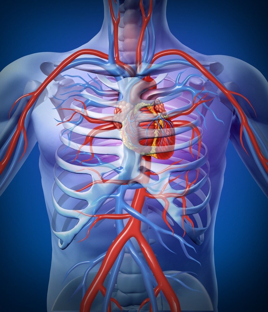
Embolization for cerebral arteriovenous malformation (AVM) treats an abnormal tangle of enlarged blood vessels, which untreated can hemorrhage (rupture and bleed) and cause a stroke. As a minimally invasive procedure, embolization for a brain AVM minimizes blood loss and allows the patient to recover more quickly than from an open brain surgery.
An embolus is an object that has circulated in the bloodstream and lodged in a blood vessel. The term embolization comes from embolus, but in this case, the embolus, in the form of a soft metal coil orbit of medical glue specially formulated for embolization of AVMs of the brain, is placed at the site of the AVM by the neurosurgeon.
The procedure treats cerebral arteriovenous malformation by using specialized neuroendovascular surgery techniques, approaches that occur inside (endo) the blood vessels (vascular) of the nervous system (neuro).
What Conditions Does Embolization for Cerebral Arteriovenous Malformation Treat?
Embolization can be used to treat a variety of conditions where blood vessels need to be occluded (closed up or blocked off) due to hemorrhaging such as gastrointestinal bleeding or to reduce the size of a tumor by cutting off the blood supply to it. However, at NSPC Brain & Spine Surgery (NSPC) (NSPC), our neuroradiologists are skilled in embolization for spine and cerebral AVMs, with specialized training in neuroendovascular techniques.
Embolization for a brain AVM can prevent a rupture, or stroke, from occurring. If bleeding has already occurred, embolization may be used to prevent further bleeding, but does not treat the post-rupture hemorrhage.
Embolization for Cerebral Arteriovenous Malformation (AVM) Treatments at NSPC
After the anesthesia is administered to the patient, a tiny incision is made at the insertion site, usually the femoral artery. A very thin, flexible tube (a catheter) is threaded from through the femoral artery up to the blood vessel in the brain with the AVM. The neuroradiologist uses fluoroscopic imaging to track and guide the catheter. Once the catheter reaches the enlarged tangle in the brain, a tiny, soft metal coil or specially formulated glue is inserted into the AVM, stopping the blood flow and preventing a hemorrhagic event. Rest after the surgery allows the insertion site into the femoral artery to heal. Usually the patient is discharged the following day. Follow up angiograms or MRAs (magnetic resonance angiograms) are usually recommended a few months after the procedure to verify the AVM embolization treatment was successful.

Physicians

Connect With Our 7 Convenient Locations
across Long Island, NY
Our expert physicians, surgeons and doctors are ready to serve you at our 7 convenient locations across Long Island, NY. Connect today to learn how our award winning, world class experts can help.
4250 Hempstead Turnpike Suite 4,
Bethpage, NY 11714
(516) 605-2720
COMMACK
353 Veterans Memorial Hwy,
Commack, NY 11725
(631) 864-3900
One Hollow Lane, Suite 212
Lake Success, NY 11042
(516) 442-2250
MANHATTAN
215 E. 77th Street Ground Floor
New York, NY 10075
(646) 809-4719
PORT JEFFERSON STATION
1500-8A Route 112,
Port Jefferson Station, NY 11776
(631) 828-3001
100 Merrick Road, Suite 128W
Rockville Centre, NY 11570
(516) 255-9031
WEST ISLIP
500 Montauk Hwy
West Islip, NY 11795
(631) 983-8400
World
Class
Expertise
For over 50 years & 350,000 patients NSPC has been a trusted global medical leader.
Contact us today for an appointment or consultation.